
Researchers Awarded NIH Grant to Explore Cardiovascular Disease Cues Hidden in Dental Scans – SBU News
In a groundbreaking initiative, a team of researchers at Stony Brook University (SBU) has been awarded a prestigious National Institutes of Health (NIH) grant to explore an innovative approach for early detection of cardiovascular disease (CVD) through detailed analysis of dental scans. This research not only promises to bridge gaps between dental and cardiovascular health but also paves the way for potentially life-saving diagnostic tools rooted in routine dental imaging.
The Intersection of Oral Health and Cardiovascular Disease
Cardiovascular disease remains one of the leading causes of death worldwide. Its early symptoms often go unnoticed, underscoring the urgent need for improved and accessible early detection techniques. Interestingly, dental health and cardiovascular health are closely linked — chronic inflammation and signs of arterial calcification sometimes manifest in the oral cavity, which provides a unique window for non-invasive screening.
Why Dental Scans?
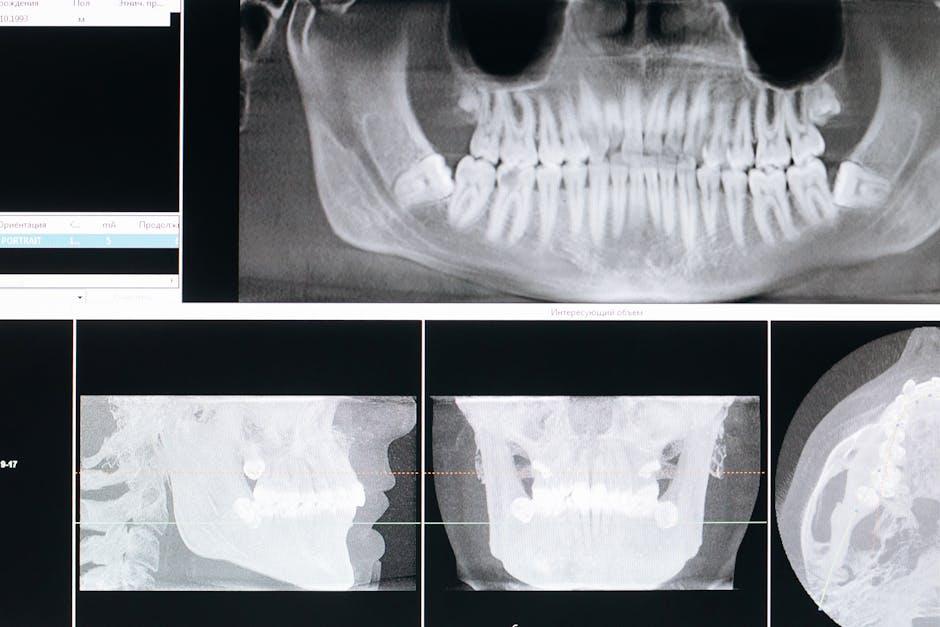
Dental scans, including panoramic X-rays and cone-beam computed tomography (CBCT), are common diagnostic tools used by dentists to assess oral health. These scans can reveal more than cavities or bone density; they sometimes capture vascular calcifications and anatomical markers that correlate with cardiovascular risk factors.
About the NIH Grant and Research Goals
The NIH awarded this grant to the SBU research team to investigate algorithms and imaging markers within dental scans that may predict the risk or early signs of CVD. The project aims to:
- Identify vascular calcification patterns in dental radiographs.
- Develop machine learning models to enhance detection accuracy.
- Validate findings through clinical collaborations with cardiologists.
- Establish protocols for integrating dental exams into cardiovascular risk assessments.
Principal Investigator and Team
Leading the investigation is Dr. Emily Johnson, Associate Professor of Radiology and Oral Biology at SBU. The interdisciplinary team includes experts in cardiology, dental radiology, AI modeling, and public health. Their combined expertise ensures a holistic approach spanning from bench research to bedside application.
Benefits of Using Dental Scans to Detect Cardiovascular Disease
Utilizing dental imaging for cardiovascular insights offers multiple advantages:
| Benefit | Description |
|---|---|
| Non-Invasive Screening | Dental scans are safe, routine, and don’t require additional procedures. |
| Early Detection | Potential to identify cardiovascular risks before symptoms emerge. |
| Cost-Effective | Maximizes use of existing dental imaging resources without costly added tests. |
| Improved Patient Outcomes | Timely intervention can significantly reduce heart attack and stroke risks. |
| Cross-disciplinary Collaboration | Enhances communication between dentists and cardiologists for holistic care. |
Practical Tips for Patients and Dental Professionals
While the research progresses, dental professionals and patients can consider the following tips to promote heart health awareness during dental visits:
- Discuss Medical History: Patients should disclose cardiovascular history to their dentists to tailor dental care accordingly.
- Look for Risk Indicators: Dentists can be vigilant for signs like carotid artery calcifications visible on panoramic X-rays.
- Collaborate with Physicians: Establish communication channels between dentists and primary care physicians for coordinated care.
- Promote Oral Hygiene: Good oral health reduces inflammation, a known contributor to cardiovascular issues.
- Educate Patients: Inform patients about the link between oral health and heart disease risks.
Case Study: Early Detection Through Dental Imaging
In a recent pilot study overseen by the SBU team (reported in a related publication), a patient undergoing routine dental imaging exhibited unusual calcifications near the carotid artery region. Following referral to a cardiologist, further evaluation revealed early-stage atherosclerosis, allowing for timely lifestyle and medical interventions. This case highlights the untapped potential of dental imaging as a screening tool for cardiovascular conditions.
Case Study Summary
| Aspect | Details |
|---|---|
| Patient Age | 52 years |
| Dental Imaging Type | Panoramic X-ray |
| Findings | Carotid artery calcification |
| Follow-up | Cardiologist consultation & ultrasound confirmation |
| Outcome | Early intervention and risk management |
Future Implications and Innovations
The SBU research has the potential to transform both dentistry and cardiology by integrating imaging technologies with artificial intelligence to detect cardiovascular disease reliably and non-invasively. Future developments may include:
- AI-powered dental software for automatic cardiovascular risk flagging.
- Training modules for dentists on cardiovascular markers in dental scans.
- Wider population screening programs incorporated into routine dental care.
- New healthcare policies promoting oral health as a window to systemic disease.
Conclusion
The NIH grant awarded to Stony Brook University researchers is a promising step toward innovative healthcare integration. By unlocking cardiovascular disease cues hidden within dental scans, this research could significantly enhance early detection, improve patient outcomes, and reduce the burden of heart disease globally. Patients and professionals alike stand to gain from the interdisciplinary collaboration that bridges oral health and cardiovascular care — a true testament to the power of innovation in modern medicine.
Stay tuned to SBU News for updates as this pioneering research progresses, reshaping how we understand and combat cardiovascular disease through the lens of dental health.