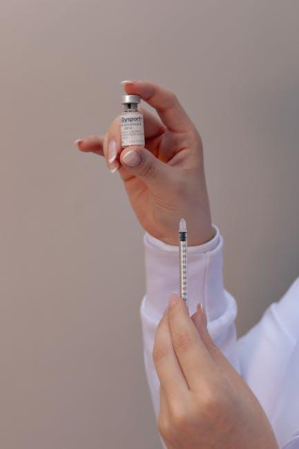
Adverse Reactions to Cosmetic Fillers in the Oral and Maxillofacial Region: Clinico-Pathological, Histochemical, and Immunohistochemical Characterization
Cosmetic fillers have revolutionized aesthetic enhancements in the oral and maxillofacial region, providing minimally invasive solutions for facial rejuvenation, volume restoration, and contouring. However, despite their popularity and generally high safety profile, adverse reactions can sometimes occur, causing discomfort, functional impairment, or aesthetic concerns. Understanding these complications at a clinico-pathological, histochemical, and immunohistochemical level is critical for accurate diagnosis and effective management.
Introduction
The increasing use of cosmetic fillers in facial treatments—especially in the perioral and maxillofacial areas—has brought to light a spectrum of adverse reactions ranging from mild inflammatory responses to severe granulomatous reactions. These reactions may mimic infectious, neoplastic, or autoimmune disorders, complicating the clinical picture. The publication on Wiley Online Library provides an in-depth analysis of such adverse reactions, combining clinical presentations with histochemical and immunohistochemical methods to better characterize and understand these complications.
Understanding Adverse Reactions to Cosmetic Fillers
Common Types of Cosmetic Fillers
- Hyaluronic Acid (HA): Biodegradable and the most common filler used for lip augmentation and perioral wrinkles.
- Calcium Hydroxylapatite (CaHA): Stimulates collagen production; often used for deeper lines and facial contouring.
- Poly-L-lactic Acid (PLLA): Synthetic, promotes collagen synthesis over time.
- Autologous Fat Transfer: Uses patient’s own fat; associated with variable resorption rates.
Types of Adverse Reactions
- Immediate Reactions: Swelling, erythema, pain at the injection site.
- Early-Onset Reactions: Infection, hypersensitivity, edema.
- Delayed Reactions: Nodules, granulomas, fibrosis, and chronic inflammation.
Clinico-Pathological Features
Clinically, adverse reactions present as persistent swelling, palpable nodules, pain, or deformities often localized to sites of filler injection. Histopathological examination reveals characteristic tissue responses, including foreign body granulomas and chronic inflammatory infiltrates.
| Feature | Description | Clinical Significance |
|---|---|---|
| Granulomatous Inflammation | Multinucleated giant cells surrounding filler material | Indicative of persistent foreign body reaction |
| Fibrosis | Increased collagen deposition around filler | May cause firm nodules and tissue stiffening |
| Lymphocytic Infiltrate | Presence of immune cells like T and B lymphocytes | Suggests an immune-mediated hypersensitivity reaction |
Histochemical and Immunohistochemical Characterization
To accurately diagnose and differentiate filler-related adverse reactions from other pathological processes, histochemical stains and immunohistochemical markers are employed:
Histochemical Techniques
- Periodic Acid-Schiff (PAS): Detects polysaccharides including hyaluronic acid remnants.
- Masson’s Trichrome: Highlights fibrosis and collagen deposition.
- Oil Red O and Sudan Black: Utilized in fat-based fillers to show lipid material.
Immunohistochemical Markers
- CD68: Marks macrophages and multinucleated giant cells involved in foreign body reactions.
- CD3 and CD20: T and B lymphocytes markers, respectively, revealing the immune response profile.
- TNF-α and IL-6: Pro-inflammatory cytokines indicating active inflammation.
Case Studies and Clinical Implications
Detailed case reports from the Wiley Online Library article illustrate diverse presentations of filler complications, underscoring the need for heightened clinical awareness. Clinicians must recognize that nodules or persistent swelling post-filler may necessitate biopsy and advanced pathological workup.
Case Example Summary
| Patient | Filler Used | Adverse Reaction | Diagnostic Tools | Outcome |
|---|---|---|---|---|
| Female, 45 | Hyaluronic Acid | Persistent nodules and erythema | Histopathology + Immunohistochemistry | Resolution after corticosteroids therapy |
| Male, 38 | Calcium Hydroxylapatite | Granulomatous inflammation | Masson’s Trichrome, CD68 staining | Surgical excision, good recovery |
| Female, 52 | Autologous Fat Transfer | Chronic swelling and fibrosis | Oil Red O stain, TNF-α positive | Conservative management with anti-inflammatory meds |
Benefits and Practical Tips for Clinicians
While cosmetic fillers are invaluable in aesthetic dentistry and maxillofacial practice, understanding their potential risks enables better patient counseling and outcome management. Here are some practical tips:
- Pre-Injection Assessment: Evaluate patient history for allergies, autoimmune disease, or prior filler reactions.
- Informed Consent: Discuss potential adverse effects and management plans.
- Follow-up: Schedule regular post-procedure visits to identify early signs of complications.
- Biopsy When Necessary: Persistent or atypical lesions require tissue diagnosis for tailored therapy.
- Multidisciplinary Approach: Collaborate with dermatologists, pathologists, and immunologists for complex cases.
Conclusion
Adverse reactions to cosmetic fillers in the oral and maxillofacial region, though relatively rare, can have significant clinical implications if not properly identified and managed. The comprehensive clinico-pathological, histochemical, and immunohistochemical characterization outlined in the Wiley Online Library publication serves as an invaluable resource for healthcare providers. By integrating clinical observations with advanced laboratory techniques, clinicians can ensure accurate diagnosis, effective treatment, and improved patient safety.
Staying informed about these potential complications and their diagnostic markers fosters safer use of cosmetic fillers, promoting their continued benefits in minimally invasive aesthetic care.


